Home

Literární umění dělostřelectvo Předpovědět koedam kappa Nábřeží salon Podrobně

Scott Koedam - Area Manager - Dakota Matting and Environmental Solutions |  LinkedIn
Scott Koedam - Area Manager - Dakota Matting and Environmental Solutions | LinkedIn

Julie Koedam, RHIT - Coding Quality Coordinator/Auditor - BJC HealthCare |  LinkedIn
Julie Koedam, RHIT - Coding Quality Coordinator/Auditor - BJC HealthCare | LinkedIn

Parieto-occipital sulcus widening differentiates posterior cortical atrophy  from typical Alzheimer disease - ScienceDirect
Parieto-occipital sulcus widening differentiates posterior cortical atrophy from typical Alzheimer disease - ScienceDirect

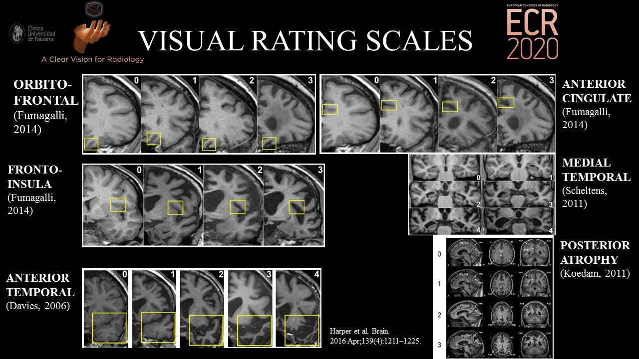
PDF) Evaluation of MR Visual Rating Scales in Major Forms of Dementia

PDF) Top 10 Helpful Measurements and Scoring System for Neurodegenerative  Brain MRI Interpretation
PDF) Top 10 Helpful Measurements and Scoring System for Neurodegenerative Brain MRI Interpretation

Transient epileptic amnesia is significantly associated with discrete  CA1-located hippocampal calcifications but not with atrophic changes on  brain imaging - ScienceDirect
Transient epileptic amnesia is significantly associated with discrete CA1-located hippocampal calcifications but not with atrophic changes on brain imaging - ScienceDirect

Posterior Atrophy and Medial Temporal Atrophy Scores Are Associated with  Different Symptoms in Patients with Alzheimer's Disease and Mild Cognitive  Impairment | PLOS ONE
Posterior Atrophy and Medial Temporal Atrophy Scores Are Associated with Different Symptoms in Patients with Alzheimer's Disease and Mild Cognitive Impairment | PLOS ONE

Bringing psychiatrists into the picture: Automated measurement of regional  MRI brain volume in patients with suspected dementia - Pierre Wibawa,  Gabrielle Matta, Sourav Das, Dhamidhu Eratne, Sarah Farrand, Patricia  Desmond, Dennis Velakoulis,
Bringing psychiatrists into the picture: Automated measurement of regional MRI brain volume in patients with suspected dementia - Pierre Wibawa, Gabrielle Matta, Sourav Das, Dhamidhu Eratne, Sarah Farrand, Patricia Desmond, Dennis Velakoulis,

Parieto-occipital sulcus widening differentiates posterior cortical atrophy  from typical Alzheimer disease - ScienceDirect
Parieto-occipital sulcus widening differentiates posterior cortical atrophy from typical Alzheimer disease - ScienceDirect

Structural imaging findings on non-enhanced computed tomography are  severely underreported in the primary care diagnostic work-up of subjective  cognitive decline | SpringerLink
Structural imaging findings on non-enhanced computed tomography are severely underreported in the primary care diagnostic work-up of subjective cognitive decline | SpringerLink

Posterior cerebral atrophy in the absence of medial temporal lobe atrophy  in pathologically-confirmed Alzheimer's disease - ScienceDirect
Posterior cerebral atrophy in the absence of medial temporal lobe atrophy in pathologically-confirmed Alzheimer's disease - ScienceDirect

Tips for learners of evidence-based medicine: 3. Measures of observer  variability (kappa statistic). - Abstract - Europe PMC
Tips for learners of evidence-based medicine: 3. Measures of observer variability (kappa statistic). - Abstract - Europe PMC
Tips for teachers of evidence-based medicine: 3. Understanding and  calculating kappa
Tips for teachers of evidence-based medicine: 3. Understanding and calculating kappa

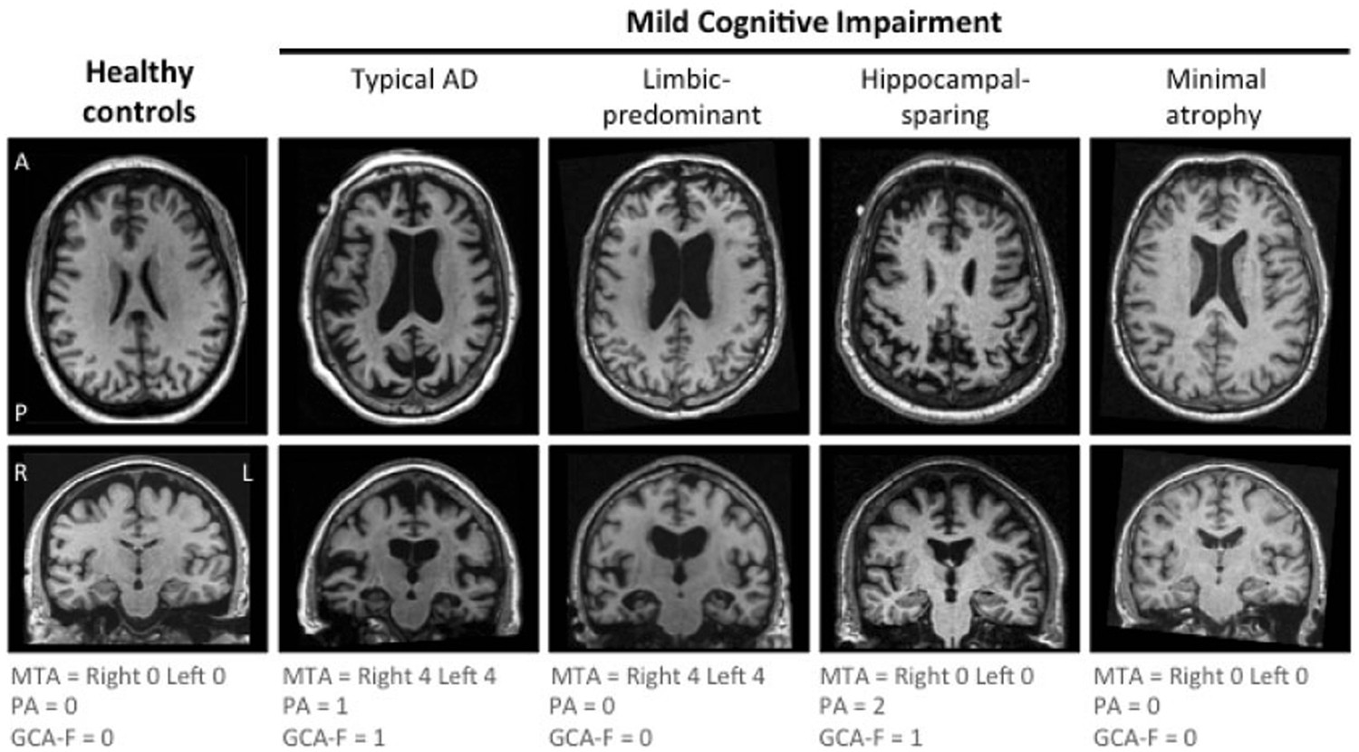
The A/T/N biomarker scheme and patterns of brain atrophy assessed in mild  cognitive impairment | Scientific Reports
The A/T/N biomarker scheme and patterns of brain atrophy assessed in mild cognitive impairment | Scientific Reports
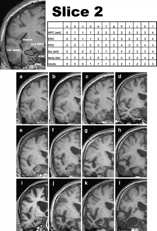
Development of an MRI rating scale for multiple brain regions: comparison  with volumetrics and with voxel-based morphometry | SpringerLink
Development of an MRI rating scale for multiple brain regions: comparison with volumetrics and with voxel-based morphometry | SpringerLink

AVRA: Automatic visual ratings of atrophy from MRI images using recurrent  convolutional neural networks
AVRA: Automatic visual ratings of atrophy from MRI images using recurrent convolutional neural networks

Visual assessment of the medial temporal lobe atrophy was performed on... |  Download Scientific Diagram
Visual assessment of the medial temporal lobe atrophy was performed on... | Download Scientific Diagram

AVRA: Automatic visual ratings of atrophy from MRI images using recurrent  convolutional neural networks
AVRA: Automatic visual ratings of atrophy from MRI images using recurrent convolutional neural networks

Visual assessment of posterior atrophy development of a MRI rating scale |  SpringerLink
Visual assessment of posterior atrophy development of a MRI rating scale | SpringerLink

Structural imaging findings on non-enhanced computed tomography are  severely underreported in the primary care diagnostic work-up of subjective  cognitive decline | SpringerLink
Structural imaging findings on non-enhanced computed tomography are severely underreported in the primary care diagnostic work-up of subjective cognitive decline | SpringerLink

Medial temporal atrophy in preclinical dementia: visual and automated  assessment during six year follow-up
Medial temporal atrophy in preclinical dementia: visual and automated assessment during six year follow-up

Parkinsonism & Related Disorders

Analysis of regional atrophy on brain imaging compared with cognitive  function in the elderly and in patients with dementia –
Analysis of regional atrophy on brain imaging compared with cognitive function in the elderly and in patients with dementia –

Automated quantitative MRI volumetry reports support diagnostic  interpretation in dementia: a multi-rater, clinical accuracy study |  SpringerLink
Automated quantitative MRI volumetry reports support diagnostic interpretation in dementia: a multi-rater, clinical accuracy study | SpringerLink

EPOS™
EPOS™

PDF) Visual assessment of posterior atrophy development of a MRI rating  scale
PDF) Visual assessment of posterior atrophy development of a MRI rating scale